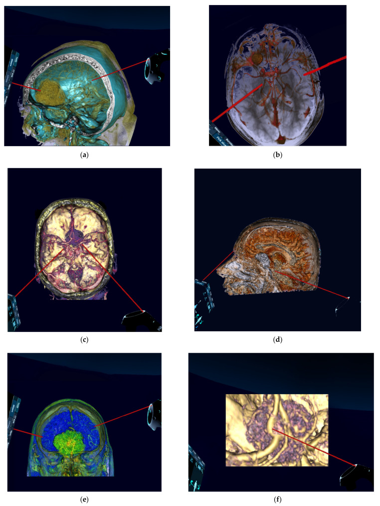
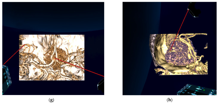
Figure 4.
Preoperative reconstructed 3D-virtual reality images (reconstructed from 2D-CT and 2D-MRI) of patients with anterior skull base meningiomas. (a) Oblique lateral aspect showing olfactory groove meningioma. (b) Superior aspect showing frontobasal meningioma. (c) Superior aspect showing planum sphenoidale meningioma. (d) Lateral aspect showing anterior clinoidal meningioma. (e) Anterior aspect showing olfactory groove meningioma. (f) Highly zoomed superior aspect showing planum sphenoidale meningioma. (g) Zoomed superior aspect showing planum sphenoidale meningioma. (h) Zoomed oblique lateral aspect showing olfactory groove meningioma. 2D, two-dimensional; 3D, three-dimensional CT, computed tomography; MRI, magnetic resonance imaging.


