Figure 5.
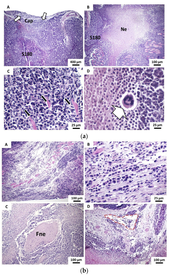
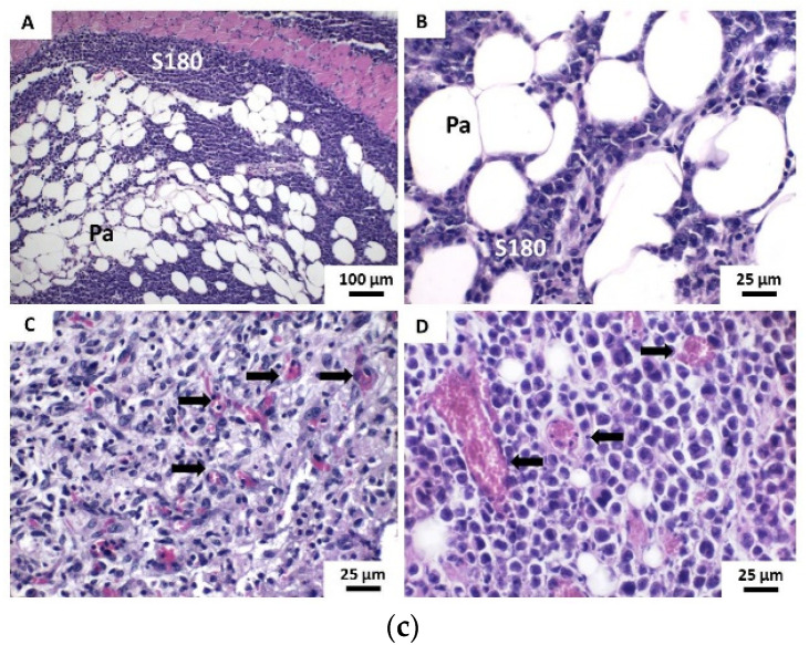
(a) Photomicrographs of histochemical technique (HE)-stained histological sections show the main histopathological features of tumors developed in the Vehicle group from the implantation of S100 cells. (A) The dermal superficial margin of the tumor shows discontinuous pseudocapsule (Cap) showing evident neoplastic infiltration (white arrows) (40×) and (B) extensive areas of coagulative necrosis (Ne) within the neoplastic cell blocks (40×). (C) Detail of tumor cells exhibit intense atypia and promote dissociation and destruction of striated skeletal muscle fibers (black arrows) (400×) and (D) Bizarre giant tumor cell (400×); (b) (A) Neoplastic cells promote infiltration and massive destruction of striated skeletal muscle fiber bundles (100×), (B) Strongly dissociated tumor cells arranged in “Indian row” cords (400×). (C) Neoplastic cells promote perineural invasion of peripheral nerve fibers (Fne) (100×). (D) Neoplastic cells observed inside the lymphatic vessel form a tumoral embolus (red outline) (100×) and (c) (A) and (B) Neoplastic cells (S180) promote massive infiltration of the hypodermic adipose panicle (Pa) (100× and 400×, respectively). (C) Intense capillary vascular neoformation (black arrows) on the periphery of the tumor and (D) in intraparenchymal areas (400×).


