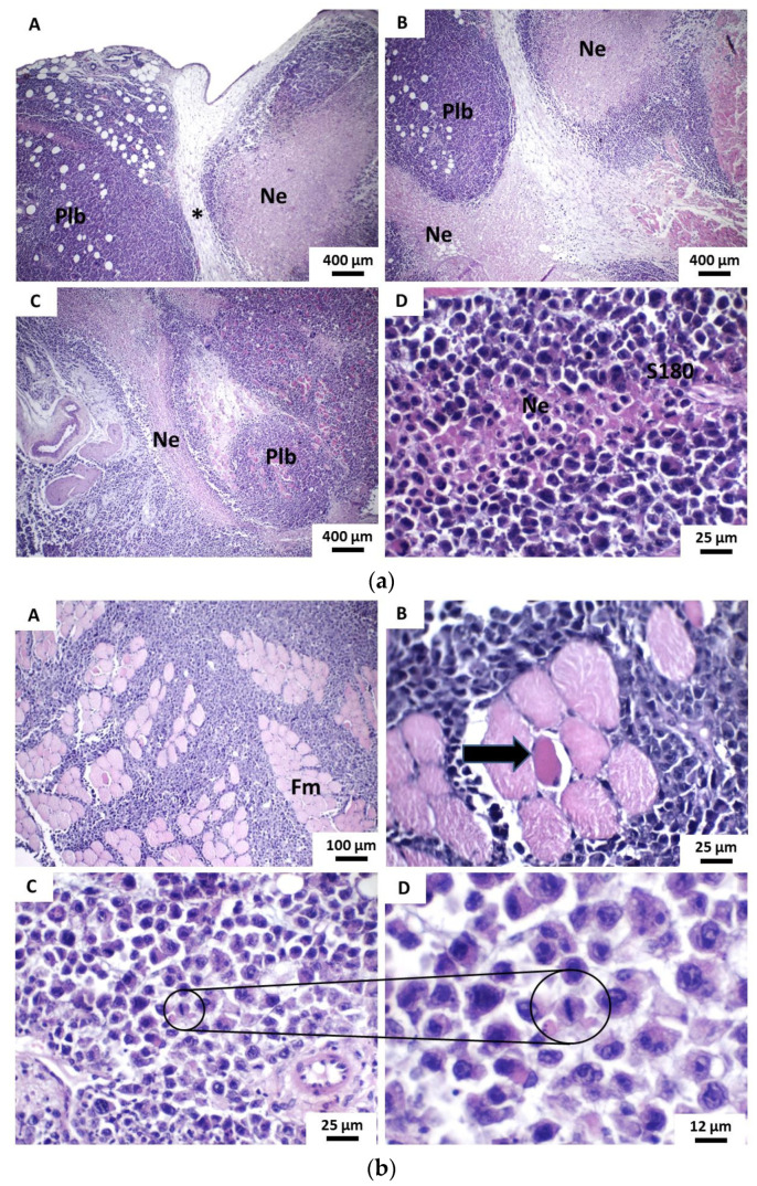
Figure 7.
Photomicrographs of histological sections stained in HE show the main histopathological features of tumors developed in the β-CD:POH 50 mg/Kg group from the implantation of S180 cells. (a) (A–C) Tumor cells organized in a pseudolobular pattern (sometimes septated by fibrous connective tissue and sometimes by irregular bands of necrotic tissue) (40×). (D) Small necrotic tissue trabic amid tumor sheet cells (400 ×). Caption: Coagulative necrosis; Plb—pseudolobules; S180—viable sarcoma 180 tumor cells and (b)(A) Tumor cells infiltrate and dissociate solid blocks of muscle fibers (Fm) (100×). (B) Apoptosis of muscle cell (black arrow) in response to tumor infiltration (400×). (C) Atypical tumor cells showing low cohesivity and mitotic activity (400×). (D) Detail mitotic figure of tumor cell in metaphasis (800×).

