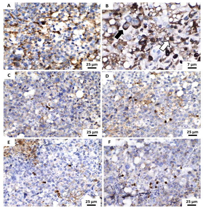
Figure 9.
Photomicrographs of histological sections demonstrate the immunohistochemical expression of the Ki67 antigen (brownish color) in the analyzed tumors. (A,B) Vehicle Group shows tumor cells showed predominantly nuclear positivity (light arrow) and eventually, cytoplasmic immunoexpression of this antigen (dark arrow). (C–F) represent the groups treated with 5-FU (25 mg/Kg/day) and 50, 100 and 200 mg/Kg/day of the formulation contain POH/β-CD inclusion complex.

