Figure 1.
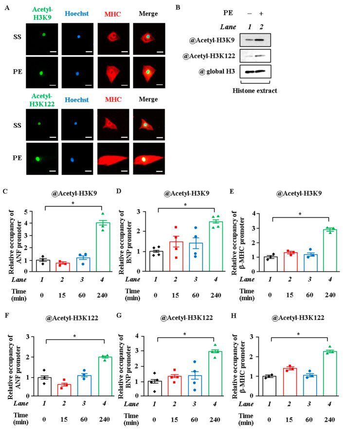
Phenylephrine stimulation induced histone acetylation in cardiomyocyte hypertrophy. (A) Primary cultured neonatal rat cardiomyocytes treated with or without phenylephrine (PE) (30 μM) for 48 h. Immunofluorescence staining was performed with anti-acetyl-histone H3K9, anti-acetyl-histone H3K122, and anti-MHC antibody. Green: Acetyl-H3K9 or Acetyl-H3K122. Blue: Hoechst. Red: MHC. Scale bar: 20 μm. (B) Histone was extracted with hydrochloric acid from cardiomyocytes treated with or without PE (30 μM) for 48 h. Western blotting was performed with anti-acetyl-histone H3K9 antibody, anti-acetyl-histone H3K122 antibody, and anti-histone H3 antibody. (C–H) ChIP assays were performed using cardiomyocyte lysates treated with or without PE for 0, 15, 60, or 240 min with anti-acetyl-histone H3K9 antibody (C–E), anti-acetyl-histone H3K122 antibody (F–H), or normal rabbit IgG as a negative control (not detected). N = 3 to 4; one-way ANOVA followed by Tukey test. * p < 0.05.

