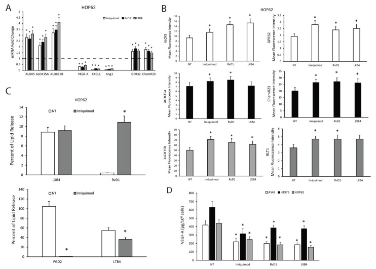
Figure 4.
Effects of TLR7 activation on SPM biosynthetic machinery and angiogenic potential of NSCLC cells. (A) ALOX5, ALOX15A, ALOX15B, VEGF-A, CXCL1, Ang1, GPR32, and ChemR23 mRNA fold change in HOP62 cells treated with imiquimod (1 μg/mL), RvD1 (1 nM), or LXB4 (1 nM) for 3 h. Data are represented as mean ± SD of five independent experiments. * p < 0.05 compared to the control (dotted line). (B) ALOX5, ALOX15A, ALOX15B, GPR32, ChemR23, and BLT1 protein expression levels (mean fluorescence intensity), assessed by cytofluorimetric analysis, in HOP62 cells treated with imiquimod (1μg/mL), RvD1 (1 nM), or LXB4 (1 nM) for 6 h. Data are represented as mean ± SD of three independent experiments. * p < 0.05 compared to the control (NT). (C) Pro-resolving and pro-inflammatory autacoids (LXB4, RvD1, PGD2, and LTB4) release in HOP62 cells treated or untreated with imiquimod (1 μg/mL, 18 h). Data are represented as mean ± SD of five independent experiments. * p < 0.05 compared to the control. (D) VEGF-A release in A549, H1975, and HOP62 cells treated with imiquimod (1 μg/mL), RvD1 (1 nM), or LXB4 (1 nM) for 10 h. Data are represented as mean ± SD of five independent experiments. * p < 0.05 compared to the control.

