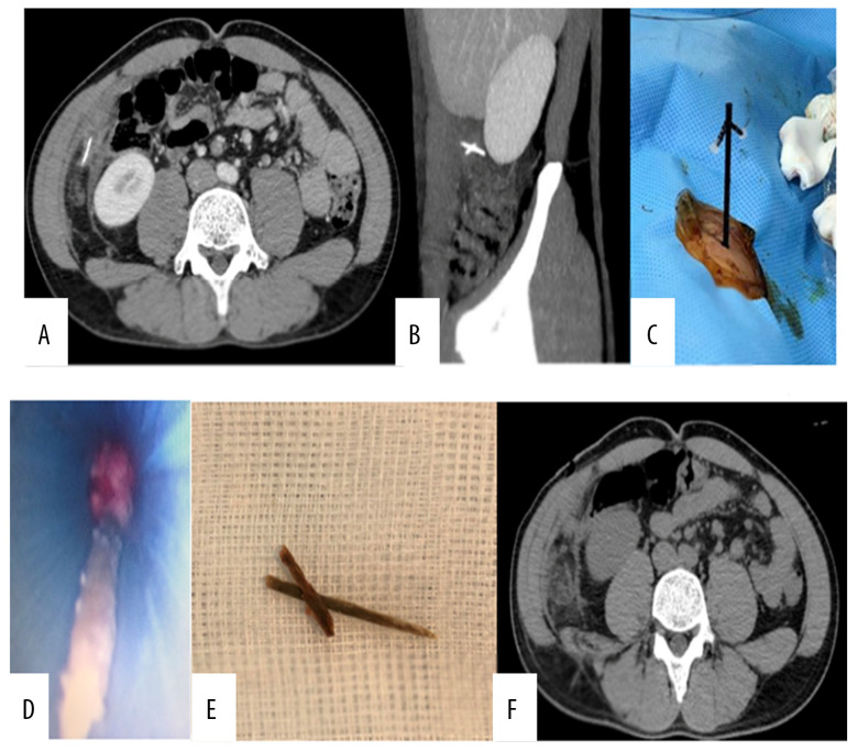
Figure 1.
(A, B) Abdominal CT scan before the intervention. The FBs appeared as hyperdensity lesions, located adjacent to each other and between the ascending colon and the right abdominal wall. (C) A plastic 14-F port was used to approach the FBs under the guidance of ultrasonography. (D) The FBs were removed through the port. (E) The FBs were 2 pieces of a wooden toothpick (11 and 21 mm). (F) The patient’s post-intervention abdominal CT scan showed that the FBs had been completely removed.

