Figure 4.
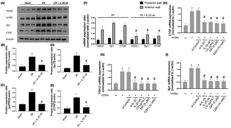
The effect of interleukin (IL)-20 antibody on ischemia/reperfusion (I/R)-induced upregulation of fibrosis factors. Sprague Dawley rats receiving sham operation or I/R treatment were studied. In the IL-20 antibody treatment group, animals were injected with 5 mg/kg IL-20 antibody at the phase of reperfusion. The left ventricular tissues were collected for Western blotting assay. Representatives of Western blot (A), and the densitometric analysis of transforming growth factor beta 1 (TGFβ1) (B), p-extracellular regulated protein kinases (ERK) (C), transcription factor specificity protein 1 (Sp1) (D), and connective tissue growth factor (CTGF) (E) were shown. (F) The mRNA levels of fibrosis factors in ischemic area (anterior ventricular wall) and nonischemic area (posterior ventricular wall) were investigated by quantitative real-time polymerase chain reaction assays. The data were presented as the mean ± standard deviation (SD) of six animals in each group. (G–I) The mRNA expression of fibrosis factors in H9C2 cells after hypoxia/reoxygenation (H/R) was examined. H9C2 cells that had been first exposed to hypoxia chamber with oxygen-glucose deprivation (OGD) for 1 h were moved into the normoxia chamber with high glucose medium for further 4 h, a condition abbreviated as H1R4. In some cases, cells were transfected with si-Control or si-IL-20 for 48 h before OGD. In the IL-20 antibody treatment group, 10 μg/mL IL-20 antibody was added to medium during H/R. Glutathione (GSH), an antioxidant, was used for mitigating oxidative stress. Diphenyleneiodonium chloride (DPI), a nicotinamide adenine dinucleotide phosphate (NADPH) oxidase inhibitor, was used to inhibit H/R-induced NADPH oxidase activation. Data were presented as the mean ± SD of three different experiments. (* indicating p < 0.05 compared to the sham group or normoxia control cells; # indicating p < 0.05 compared to the I/R group or H/R cells).

