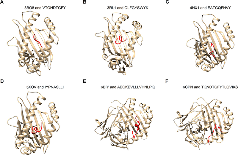
Figure 5: Docking of epitopes to HLA structures.
The epitopes (red) were docked to six HLA 3D structures available in PDB using Boston University’s ClusPro server. The six complex structures found in PDB include four MHC Class I molecules (A-D, see Table 1) and two MHC Class II molecules (E-F, see Table 2). The epitopes were modeled by PEP-Fold 3.0 that uses ab initio modeling.

