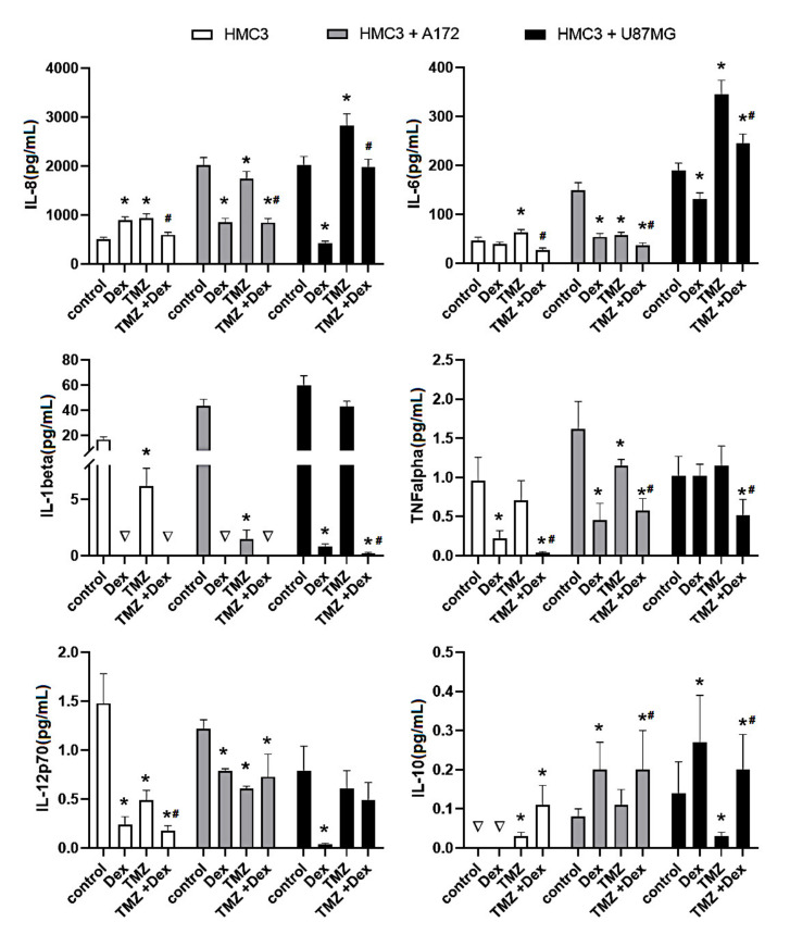
Figure 1.
Flow cytometric analysis of population of HMC3 cells for expression of IL-8, IL-6, IL-1β, TNFα, IL-12p70 and IL-10. Cells were grown in monoculture or co-culture and exposed to Dex (10 μM) and/or TMZ (10 μM) for 24 h. The immune cell samples were collected and stained with phycoerythrin-conjugated antibodies for expression of selected cytokines. The concentration of the target proteins was determined using the standard curve according to the manufacturer instructions. * p < 0.05 vs. corresponding control group, # p < 0.05 TMZ + Dex vs. TMZ treated group; ∇—concentration <0.01 pg/mL. Control—nonstimulated cells, Dex—dexamethasone alone treated cells, TMZ—temozolomide alone treated cells, TMZ + Dex—cells treated with temozolomide in combination with dexamethasone.

