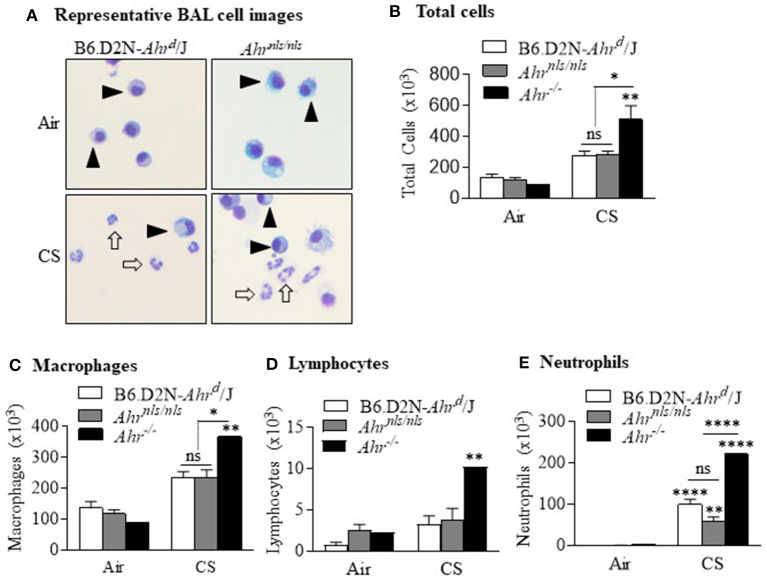
Figure 3.
Suppression of acute smoke-induced neutrophilia does not require nuclear localization of the AhR. (A) Most cells within the BAL of air exposed B6.D2N-Ahrd/J and Ahrnls/nls mice were macrophages (arrowheads). There was a noticeable increase in neutrophils after CS exposure (open arrows). Representative images are shown. (B) There was an increase in the total number of BAL cells after CS exposure (**p < 0.01). There was also a significant difference in total cells between the CS-exposed Ahr−/− mice and both B6.D2N-Ahrd/J and Ahrnls/nls mice (*p < 0.05) but not between B6.D2N-Ahrd/J and Ahrnls/nls mice (ns). (C) There was an increase in the number of macrophages in CS-exposed Ahr−/− mice. (D) There was no change in the number of lymphocytes. (E) There was a significant increase in the number of neutrophils after acute CS exposure (**p < 0.01; ****p < 0.0001 compared to respective air group). There was a stronger neutrophilic response in the Ahr−/− mice compared to the CS-exposed B6.D2N-Ahrd/J and Ahrnls/nls mice (****p < 0.0001). There was no difference in neutrophils between the CS-exposed B6.D2N-Ahrd/J and Ahrnls/nls mice. Results are shown as means ± SEM (n = 5–12 male and female mice per group).

