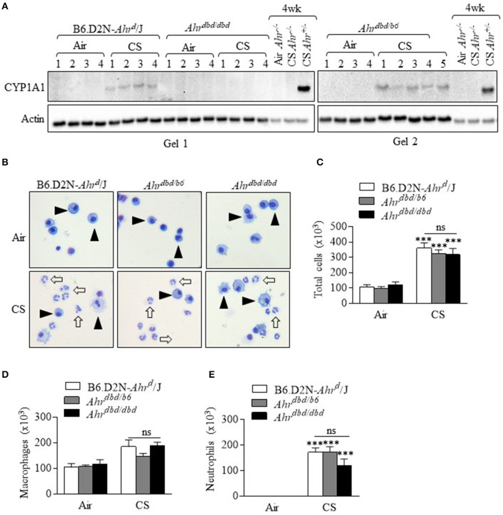
Figure 4.
The AhR suppresses acute CS-induced neutrophilia independent of the DRE. (A) CS exposure for 3 days robustly increased CYP1A1 in the control mice (B6.D2N-AhRd/J- Gel 1 and Gel 2. There was no induction in CYP1A1 expression in Ahrdbd/dbd mice (Gel 1). Additional controls included Ahr−/− mice exposed to CS for 4 weeks (no CYP1A1) and strong CYP1A1 protein in Ahr+/− mice. Numbers represent individual mice. (B) Macrophages were the predominant cell type in the BAL in the air exposed mice (arrowheads). Acute CS exposure noticeably increased the abundance of neutrophils (open arrows). Representative images are shown. (C) There was a significant increase in the total number of BAL cells from CS exposure (***p < 0.0001, CS vs. air-exposed mice). There was no significant difference (ns) in the total number of cells between the B6.D2N-Ahrd/J, Ahrdbd/b6, and Ahrdbd/dbd mice. (D) There was no significant difference (ns) in the number of macrophages between the B6.D2N-Ahrd/J, Ahrdbd/b6 and Ahrdbd/dbd mice. (E) CS exposure significantly increased the number of neutrophils in the BAL in each group (***p < 0.001 compared to air-exposed mice). There was no significant difference (ns) in the total number of neutrophils between the B6.D2N-Ahrd/J, Ahrdbd/b6 and Ahrdbd/dbd mice exposed to CS. Results are shown as means ± SEM (n = 4–5 female mice per group).

