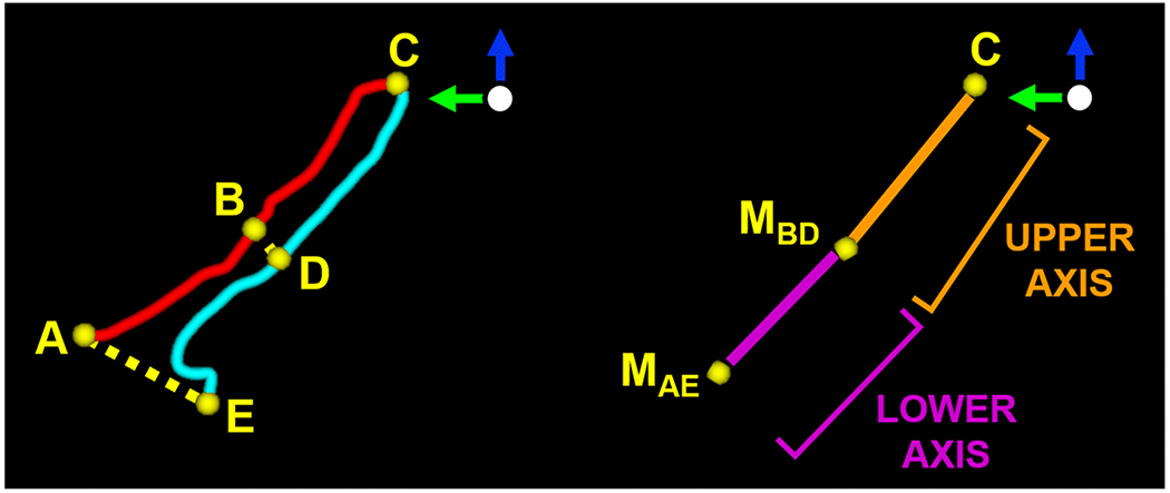
Figure 5.

Visualization of the vaginal position and orientation with respect to the Y (green) and Z axes (blue) of the 3D pelvic coordinate system. Left: anterior hymenal remnant (point A), halfway point of the anterior vaginal wall (point B), vaginal apex (point C), halfway point of the posterior vaginal wall (point D), and posterior hymenal remnant (point E) are identified along the vaginal contour. The distance between the hymenal remnants (dotted line AE) and halfway points (dotted line BD) are also displayed. Right: vaginal apex (point C) and midpoints of the i) hymenal remnants (point MAE) and ii) halfway marks (point MBD) delineate the upper axis (orange line) and lower axis (purple line) of the vagina.
