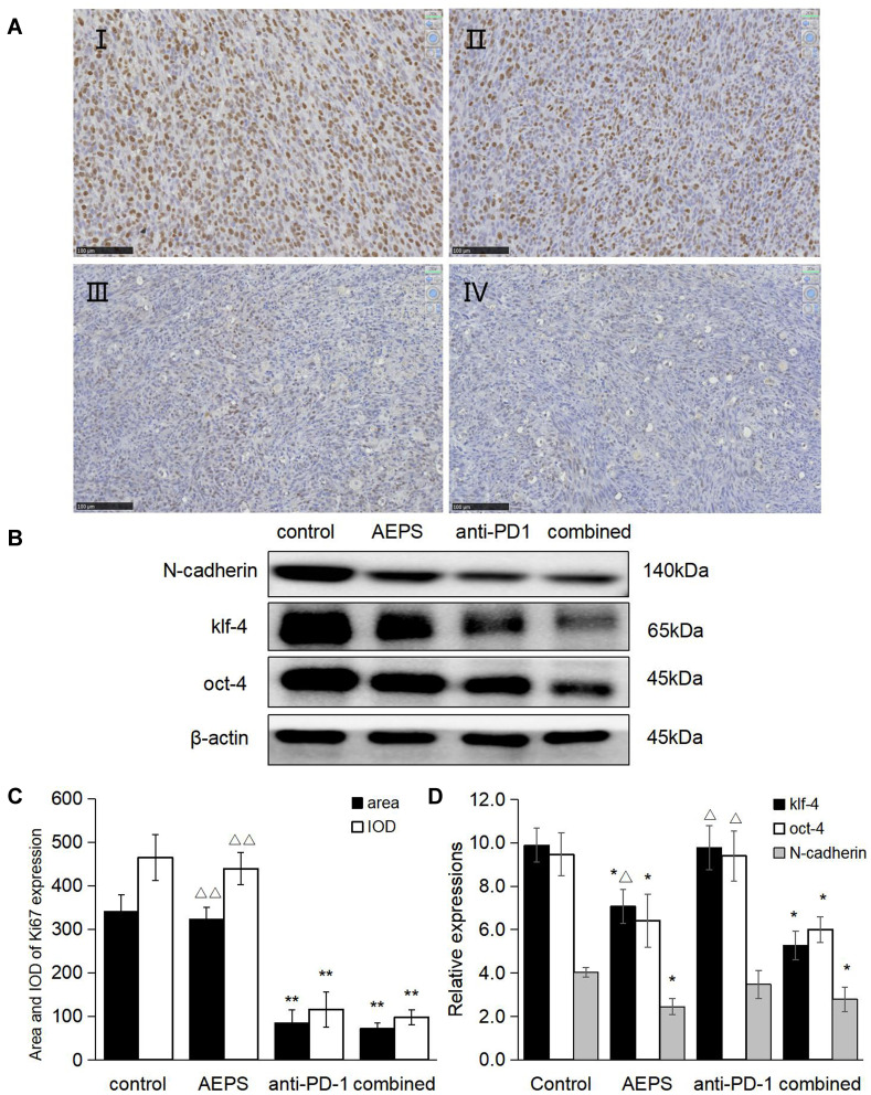
Figure 4.
Combination of AEPS and PD1 antibody inhibited tumor proliferation and metastasis-marker protein expression in colorectal cancer–xenograft mice. CT26 cells were inoculated to establish the colorectal cancer–xenograft mouse model, 5 mL/kg•day AEPS given orally, and 10 mg/kg•week anti-PD1 injected intravenously separately or combined for 28 days. (A) After 28 days, tumors in each group were removed and sliced, IHC staining performed with the anti-Ki67 primary antibody, and slices observed and photographed under 200× magnification. (I) control group, (II) AEPS group, (III) anti-PD1 group, (IV) combined group. (B) Western blot was performed with anti-N-cadherin, anti-KLF4, and anti-Oct4 primary antibodies. (C) Area and integral optical density of Ki67 was analyzed with Image-Pro Plus 6.0. (D) Semiquantification of protein levels was performed with ImageJ software, and relative expressions of proteins calculated using β-actin as an internal control. Statistical analysis was performed using Student’s t-test. *P<0.05, **P<0.01 compared to the control group; ΔP<0.05, ΔΔP<0.01 compared to the combined group.

