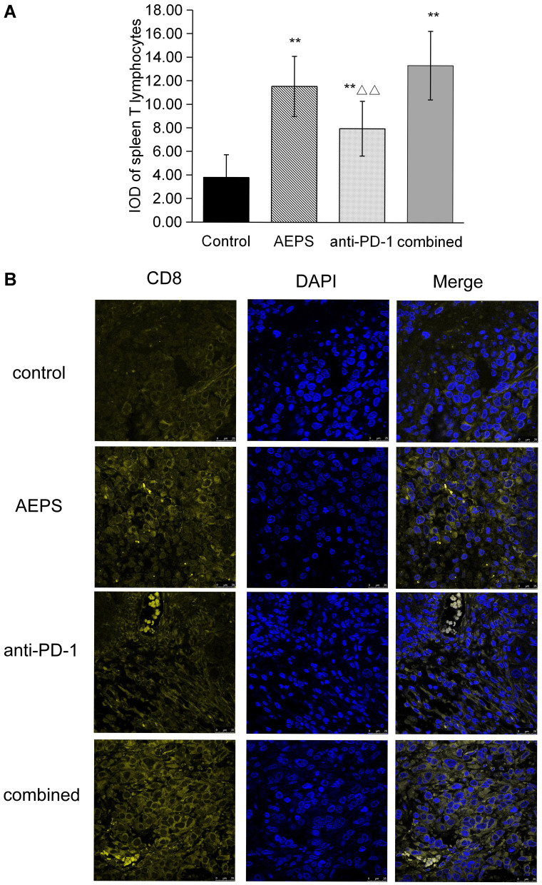
Figure 5.
Combination of AEPS and PD1 antibody stimulated spleen T-cell proliferation and tumor CD8+ T-cell infiltration in colorectal cancer–xenograft mice. CT26 cells were inoculated to establish the colorectal cancer–xenograft mouse model, 5 mL/kg•day AEPS was given orally, and 10 mg/kg•week anti-PD1 injected intravenously separately or combined for 28 days. (A) After 28 days, spleens were removed and cells extracted and cultured. ConA was used to induce the proliferation of spleen lymphocytes, proliferation activity of T cells was detected with MTT, and optical density measured at 570 nm wavelength. Statistical analysis was performed using Student’s t-test. **P<0.01compared to the control group; ΔΔP<0.01 compared to the combined group. (B) Immunofluorescence analysis was performed with anti-CD8α primary antibody, coverslipped slides were covered with DAPI, and merged images show the overlap of yellow CD8 and blue DAPI fluorescence.

