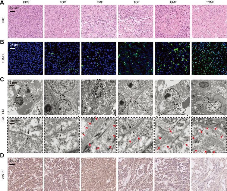
Figure 7.
(A) He staining was used to detect the morphological changes of HeLa tumor after receiving PBS, TGM, TMF, TGF, GMF and TGMF treatment. (B) TUNEL staining was used to detect the proportion of apoptotic cells in HeLa tumor after receiving PBS, TGM, TMF, TGF, GMF and TGMF treatment. (C) The presence of NFS in HeLa tumor cells was detected by bio-TEM (the black arrow is cell membrane, and the red arrow is NFs). (D) The expression of WNT1 in HeLa tumor was detected by IHC staining.

