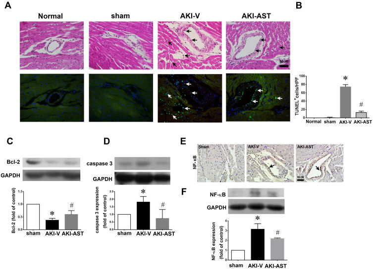
Figure 7.
AST-120 therapy reduced cell apoptosis and NF-κB activation following renal I/R injury. (A and B) Detection of apoptosis (arrows) by HE staining (upper panels) and TUNEL staining (lower panels) in the normal (both kidneys intact), sham, AKI+V, and AKI+AST groups. The number of apoptotic cells per high-power field was counted. n = 4 per group; the data are presented as mean ± SEM, *p < 0.05 vs the sham group; #p < 0.05 vs the AKI+V group. (C and D) The expression of Bcl-2 and caspase 3 at day 2 after I/R injury in heart tissues from the sham, AKI-V, and AKI-AST groups determined by Western blotting. n = 6 per group; the data are presented as mean ± SEM, *p < 0.05 vs the sham group, #p < 0.05 vs the AKI+V group. (E and F) NF-κB expression in the three groups was screened using an immunohistochemistry assay. Representative photomicrographs of immunohistochemical staining for NF-κB in heart tissues from the sham, AKI-V, and AKI-AST groups. n = 4; the data are presented as mean ± SEM, *p < 0.05 vs the sham group; #p < 0.05 vs the AKI-V group.

