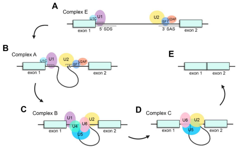
Figure 2.
Schematic representation of the splicing process. (A) Assembly of the spliceosome: U1 snRNP recognizes the splice donor site (SDS) and U2 snRNP recognizes the splice acceptor site (SAS) to generate complex E. (B) U2 recognizes the adenosine at the branch-site and forms complex A. (C) The U4/U6·U5 tri-snRNP joins the spliceosome to form complex B. (D) U4 and U1 are released, U6 replaces U1 recognizing the SDS and interacts with U2, generating complex C and catalyzing the splicing reaction. (E) Exons are ligated, and intronic pre-mRNA and spliceosomal snRNPs are liberated.

