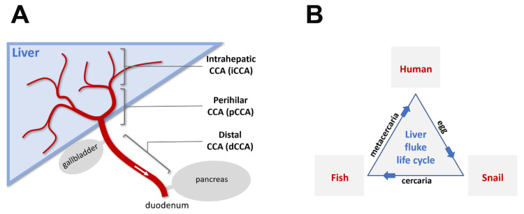
Figure 1.
Schematic diagram of cholangiocarcinoma (CCA) types and the liver fluke life cycle. (A): The bile duct and its relationship to the liver, gallbladder, pancreas, and duodenum. The biliary tree (red) is composed of the ampulla of Vater (opening to the duodenum part of the small intestine after joining of the pancreatic duct), common bile duct (after joining of the cystic duct from gallbladder, before joining of the pancreatic duct), common hepatic duct (after joining of the left and right hepatic ducts, before joining of the cystic duct), left and right hepatic ducts, and progressively narrower intrahepatic ducts and ductules. CCAs are categorized based on the origin of tumorigenic cholangiocytes within the biliary tree. Fluke-induced CCAs are primarily of the intrahepatic type, likely due to comparable lumen diameters with respect to the size of adult parasitic flukes. (B): Life cycle of liver flukes in humans. Parasitic liver flukes (Opisthorchis viverrini and Clonorchis sinensis) use the human bile duct as a host environment for sexual maturation. Eggs generated from cross-fertilization of hermaphroditic adult liver flukes pass through feces and infect the first intermediate host (snails) after a brief period of free-living development. Asexual reproduction takes place in this host environment. Larval flukes (cercariae) released from infected snails metamorphose into encysted metacercariae either in a natural environment or in a second intermediate host (freshwater fish for liver flukes in humans). The encystment in fish muscular tissues presumably confers evolutionary advantage for liver flukes in reaching their final parasitic host environment, where they can live for up to two decades.

