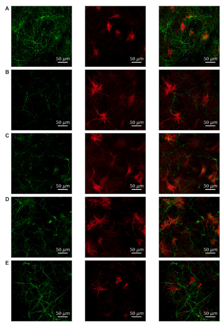
Figure 3.
Morphology of primary hippocampal cultures on day 21 of cultivation in vitro. Green, stained by a marker of neuronal protein (MAP2); red, marker of cytoskeleton protein of differentiated astrocytes (GFAP). (A) Sham. (B) Hypoxia. (C) Glucose deprivation. (D) Hypoxia + FLT4 blocker. (E) Glucose deprivation + FLT4 blocker. Scale bars: 50 µm.

