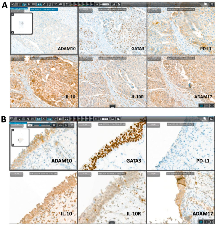
Figure 1.
Representative immunohistochemical (IHC) figures of the complete IHC panel stained on consecutive sections using CaseViewer digital microscopy for systematic analysis. The presented cases show a pT2b urothelial carcinoma (A) and a primary carcinoma in situ (CIS) (B) on radial cystectomy (RC) specimen.

