Figure 4.
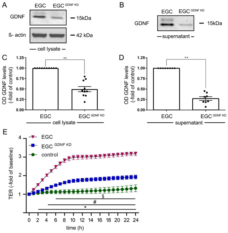
Knockdown of glial cell line-derived neurotrophic factor (GDNF) reduced effects of enteric glial cells (EGC) on barrier properties of Caco2 cells. (A). Representative Western blot of cell lysates of EGC to confirm reduced GDNF levels following knockdown of GDNF in EGC (EGCGDNF KD); (n = 10). (B). Representative Western blots of cell culture supernatants from EGCs and EGCGDNF KD to confirm reduced secretion of EGCs into cell culture supernatants; (n = 8). (C). Quantifications of Western blots following knockdown of GDNF showed reduced levels to 45 ± 19% of control EGCs correlated to β-actin; (n = 10, ** = p < 0.01, Wilcoxon signed-ranked test). Control = EGC WT. (D). Quantifications of Western blots from supernatants from EGC supernatants compared to EGCGDNF KD demonstrated reduced GDNF levels of 27 ± 12%; (n = 8, ** = p < 0.01, Wilcoxon signed-ranked test). Control = EGC WT. (E). Measurements of TER revealed that incubation of Caco2 with EGC supernatant significantly increased TER to 330% ± 16% after 24 h whereas incubation with supernatants from EGCGDNF KD resulted in a significantly less pronounced increase of TER to 230% ± 18% (equates to 0.69-fold of EGC supernatants); (n = 10 for each condition, § = p< 0.05 control vs. EGC, # = p < 0.05 EGC vs. EGCGDNF KD, * = p < 0.05 control EGC; two-way ANOVA); Control = Caco2 monolayer without treatment.

