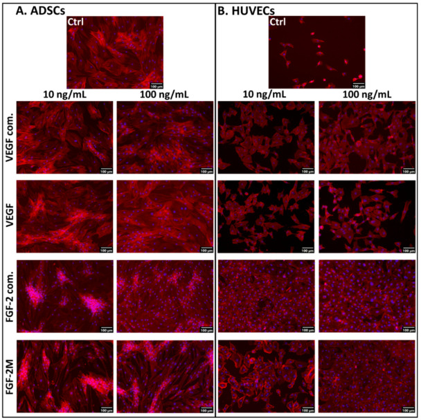
Figure 2.
Microphotographs of ADSCs (A) and HUVECs (B) on day 7 after seeding in media enriched with commercial VEGF-A165 (VEGF com.) or our recombinant VEGF-A165, and in media enriched with commercial FGF-2 (FGF-2 com.) or our recombinant FGF-2M. The growth factors were added into DMEM with 10% FBS for ADSCs and into EGM2-weak for HUVECs. Representative low and high concentrations (i.e., 10 ng/mL and 100 ng/mL, respectively) of the tested growth factors were selected. Control cells were grown in media without growth factors (Ctrl). The filamentous actin in cells was stained with phalloidin-tetramethylrhodamine (TRITC) to visualize the cell morphology. The nuclei were counterstained with Hoechst 33258. Olympus IX 71 microscope, DP 70 digital camera, obj. 10×, scale bar 100 μm.

