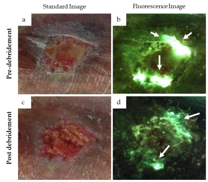
Figure 4.
Presence of cyan fluorescence in a 78-year-old female patient with a venous leg ulcer. Standard images (a,c) and fluorescence images (b,d) were captured at the point-of-care before and after debridement. Bright white/cyan fluorescence signal was observed before debridement (b). Cyan fluorescence signal intensity was significantly reduced after debridement (d) but was still clearly present (white arrows).

