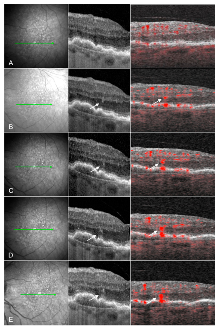
Figure 2.
Courtesy of Sacconi et al. 2019: Evolution of type 3 macular neovascularization: Near infrared (NIR) reflectance (left column), structural optical coherence tomography (OCT) B-scan (middle column), and cross-sectional OCT angiography (OCTA) (right column) images of one eye with a type 3 macular neovascularization (MNV) from a patient with age-related macular degeneration (AMD) at (A) the first preclinical stage examination and after (B) 5 months, (C) 8 months, (D) 12 months, and (E) 15 months. The MNV originates from the deep retinal capillary plexus, evident as a small intraretinal hyperreflective focus on OCT and a punctate flow signal on cross sectional OCTA (white arrows). The lesion progresses downward toward the RPE over time (from (A) to (E)). These lesions may be driven by choriocapillaris ischemia, as eyes with type 3 MNV have significantly increased choriocapillaris flow deficits with OCTA [95,96].

