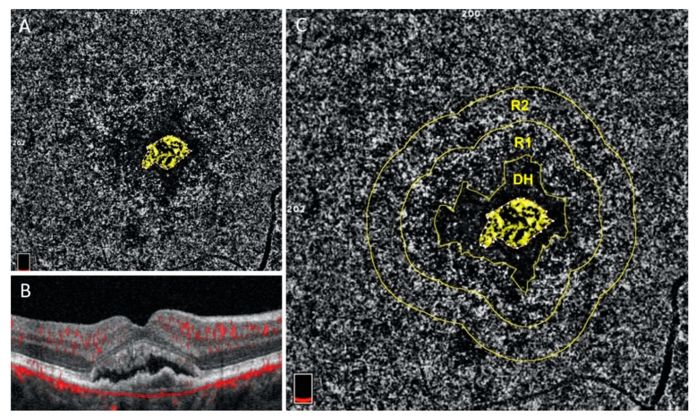
Figure 4.
Courtesy of Scharf et al. 2020: Choriocapillaris flow deficits around macular neovascularization: En face choriocapillaris OCTA (A and C) and structural OCT B-scan (B) images of treatment-naïve exudative macular neovascularization (MNV). In the Scharf study, en face choriocapillaris angiograms were analyzed for the percentage of choriocapillaris (CC) flow deficits in two concentric rings, (R1) and (R2), around the peri-lesional dark halo (DH), (image C). The ring closer to the MNV (R1) exhibits significantly greater percentage of flow deficits than the more peripheral ring. Both rings exhibit significantly greater flow deficits than the same areas in age-matched normal controls [44].

