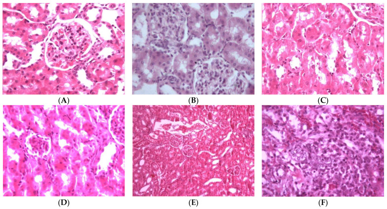
Figure 7.
Light microscope pictures of kidney cells: (A) Normal cells. (B) Kidney cells of Pa-treated group showing abnormal cellular structures with degenerated corpuscle, reduced size of glomeruli, shrinkage of Bowman capsule, cloudy swelling in tubules and inflammatory infiltrate. (C) Kidney cells of rats treated with Pa and Sil showing no marked changes, nearly normal glomeruli and mild cloudy swelling in tubules. (D) Kidney cells of group treated with 3 showing moderate cortical atrophy and reduced size of glomeruli. (E) Kidney cells of group treated with 4 showing kidney cortical congestion, abnormal glomeruli (reduced in size) and cloudy swelling in tubules. (F) Kidney cells of group treated with 6 showing inflammatory cell infiltration at corticomedullary junction, kidney cortical infiltration, almost complete absence of Bowman space, degenerated and convoluted necrotic tubules.

