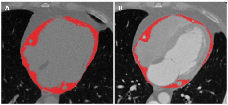
Figure 1.
Segmentation of epicardial adipose tissue with application of thresholding, thus only including voxels with a density values between −190 and −30 HU, on both the unenhanced (A) and contrast-enhanced (B) scans. The paracardiac adipose tissue lies right outside the pericardium, surrounding the epicardial adipose tissue.

