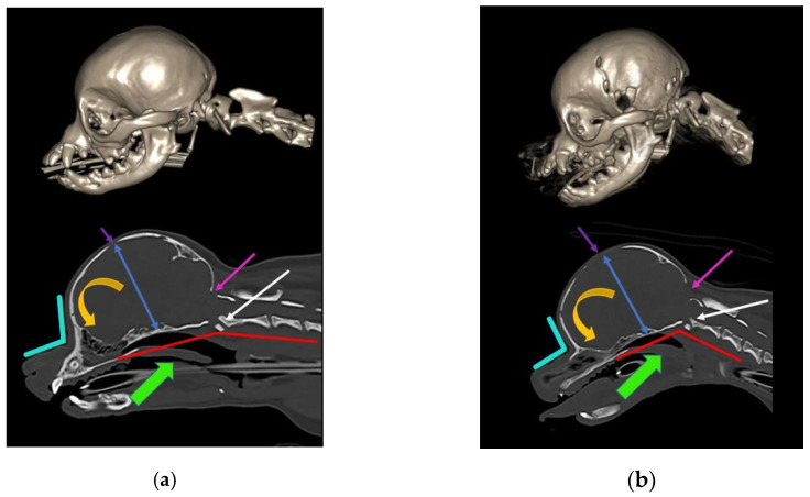
Figure 1.
Skull and craniocervical junction changes with extreme brachycephaly. Reconstructed and midsagittal CT of two female sibling Chihuahuas aged one year old. (a) Less extreme 2.5 kg female Chihuahua, sibling to (b). (b) More extreme miniaturized and brachycephalic 1.6 kg female Chihuahua, sibling to (a). With increasing brachycephaly, the angle of stop (junction between frontal and nasal bones; aqua angle) becomes more acute. With more extreme brachycephaly, ventral brain rotation is more pronounced (orange arrow). There is a relative increase in the cranium height (blue arrow), and the molera (persistent bregmatic fontanelle; purple arrow) is wider. Altered occipital bone conformation changes the angle between the skull base and the cervical vertebrae, resulting in a cervical flexure (red line) and dorsal tipping of the odontoid peg into the spinal cord (white arrow). The oropharynx is displaced caudally (green arrow). There is also a Chiari-like malformation with a small caudal fossa, reduced occipital crest, and a short, more vertical supraoccipital bone (pink arrow). The supraoccipital bone has failed to ossify ventrally. The atlas is closer to the skull, contributing to the craniocervical junction overcrowding (images created by C. Rusbridge and S.P. Knowler).

