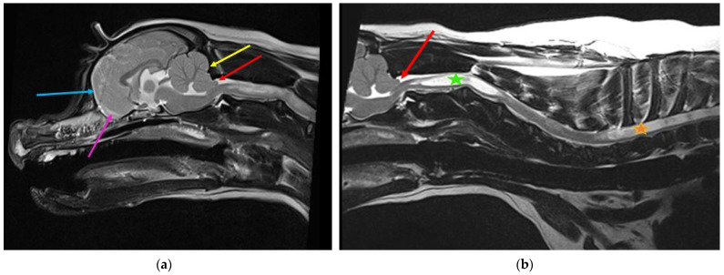
Figure 5.
Two-year-old female Cavalier King Charles spaniel with Chiari-like malformation and syringomyelia. (a) T2-weighted mid-sagittal brain MRI. There is rostrotentorial crowding, giving the rostral forebrain a flattened appearance (blue arrow), with reduced size and ventrally displaced olfactory bulbs (pink arrow). The caudal fossa is reduced rostrally by the displaced forebrain and caudally by a short vertical supraoccipital bone (yellow arrow). The cerebellum is flattened against the supraoccipital bone, resulting in caudal vermal indentation and herniation into or through the foramen magnum (red arrow). (b) T2-weighted mid-sagittal cervicothoracic spinal MRI showing cerebellar vermis herniation (red arrow). There is wide syringomyelia in the cervical (green star) and thoracic (orange star) spinal cord. This dog was presented with signs of pain and fictive scratching (images created by C. Rusbridge and S.P. Knowler).

