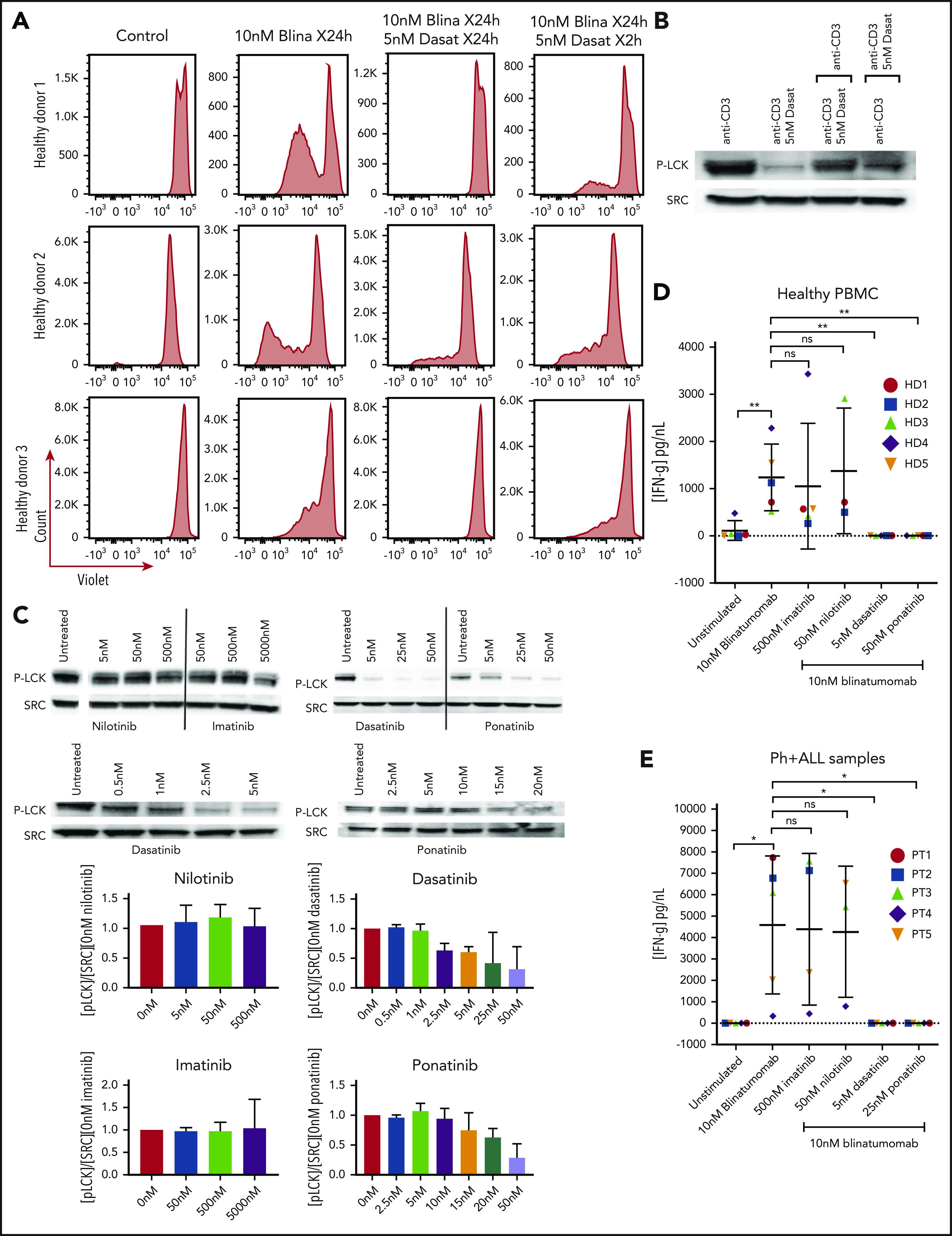
Figure 2.

Assessment of brief exposure, cytokine release, and activation of LCK. (A) PBMC response to brief treatment of dasatinib from 3 healthy donors. Cells from healthy donors were cultured for 5 days alone or with 10 nM blinatumomab with 5 nM dasatinib for 24 hours compared with 2 hours. Cells were then assessed by multiparametric flow cytometry gated for CD45+ cells. Histograms of CD3+ cells were assessed for retention of CellTrace Violet. Loss of intensity is a marker of cell proliferation. (B) Recovery of the T-cell response after dasatinib treatment. Jurkat cells were cultured with anti-CD3 and anti-CD28, or anti-CD3 and anti-CD28 plus dasatinib for 2 hours. After 2 hours, anti-CD3–treated cells were treated with anti-CD3 plus dasatinib, and the CD3 plus dasatinib treated cells were treated with anti-CD3 only. Cell lysate was then isolated, separated, and immunoblotted for phospho-tyr 394 LCK (P-LCK) or total Src. (C) Assessment of LCK phosphorylation by treatment with ABL kinase inhibitors. Jurkat cells were stimulated at baseline with CD3 and CD28 for 4 hours without (untreated) or with nilotinib, imatinib, dasatinib, and ponatinib at the specified concentration. Cell lysate was then isolated, separated, and immunoblotted for phospho-tyr 394 LCK (P-LCK) or total Src. P-LCK blots were quantified compared with total SRC and then normalized to untreated. Histograms below the blots represent the percent inhibition with the standard deviation. (D) IFN-γ expression from PBMCs from healthy donors. Samples were either unstimulated or treated with 10 nM blinatumomab with or without 500 nM imatinib, 50 nM nilotinib, 5 nM dasatinib, or 25 nM ponatinib for 5 days. Media were then harvested and assessed for concentration of IFN-γ (IFN-γ). Comparisons were performed by Student t test (*P < .05, **P < .01). (E) IFN-γ expression from Ph+ ALL samples. Samples were either unstimulated or or treated with 10 nM blinatumomab with or without 500 nM imatinib, 50 nM nilotinib, 5 nM dasatinib or 25 nM ponatinib for 5 days. Media were then harvested and assessed for concentration of IFN-γ. Comparisons were performed bu Student t test (*P < .05, **P < .01).
