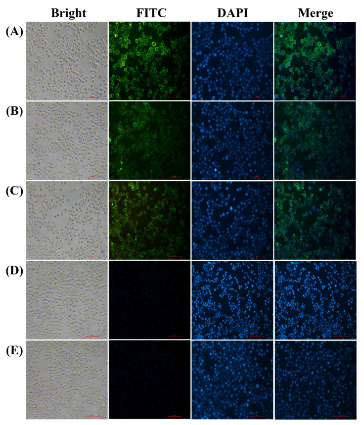
Figure 1.
Immunofluorescent staining analysis of M, S and E protein expression in the recombinant baculovirus-infected Sf9 cells at 60 h post-infection (200×). (A) Sf9 cells infected with recombinant baculovirus rHBM-S. (B) Sf9 cells infected with recombinant baculovirus rHBM-M. (C) Sf9 cells infected with recombinant baculovirus rHBM-E. (D) Sf9 cells infected with wild-type baculovirus. (E) Sf9 cells. FITC was antibody conjugated (green); DAPI was used to stain cell nuclei (blue), and merging signified that FITC merged with DAPI.

