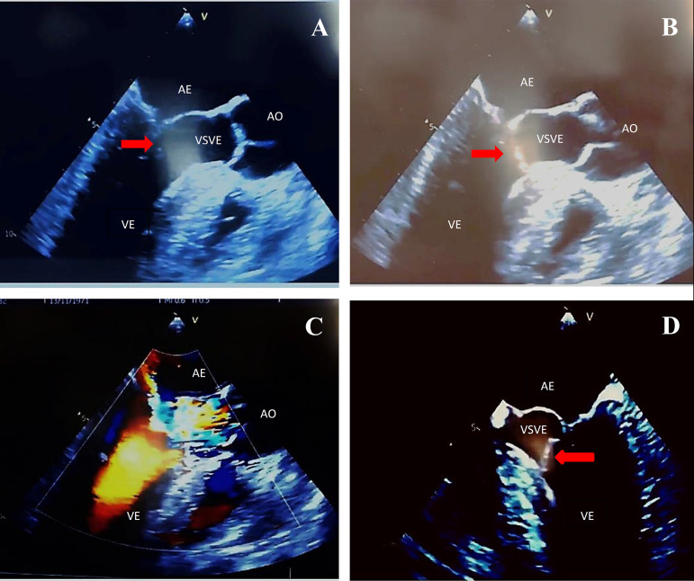
Fig. 1.
(A) Mitral valve partially closed with hyperechogenic image (red arrow) adhered to its anterior leaflet (esophageal image with long axis at 120 degrees); (B) mitral valve during ventricular systole with a hyperechogenic image (red arrow) adhered to its anterior leaflet, partially obstructing the LVOT (120-degree long-axis esophageal image); (C) acceleration of systolic flow due to partial obstruction of the LVOT (long-axis esophageal image at 120 degrees); (D) hyperechogenic image (red arrow) adhered to the anterior mitral valve cusp partially obstructing LVOT in systole (esophageal image of 4 chambers at 0 degrees). AO=aorta; LA=left atrium; LV=left ventricle; LVOT=left ventricular outflow tract

