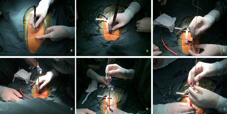
Fig. 1.
Images of the procedure. A: planning the incision site. B: selection of the puncture site in the right ventricle. C: an 18G venous indwelling needle was inserted through a purse-string suture, and a guidewire was advanced to the right ventricle. D: the needle was removed, and a delivery sheath was advanced over the guidewire. E: the device was advanced through the delivery sheath. F: the device was deployed after the device was in the correct position.

