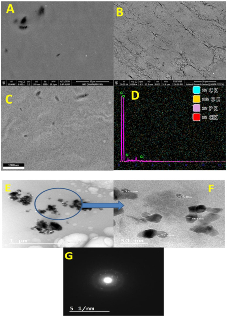
Figure 3.

Scanning electron microscope topography of glycogen (A), gelatin (B), green nanocomposite-based biopolymers (Ge-Nco) (C), and mapping as well as energy dispersive X-ray chart (D). Transmission electron microscope of green nanocomposite-based biopolymers at low magnification (E), at high magnification (F), and diffraction of nanoparticle (G).
