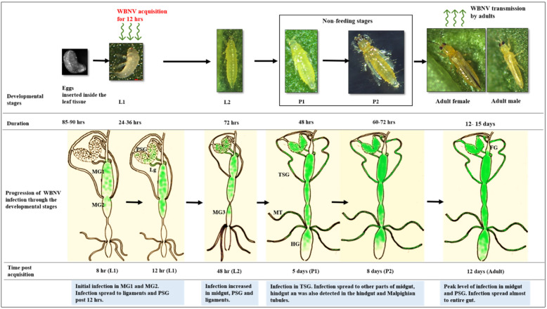
Figure 10.
A schematic representation of WBNV dynamics from egg to adult stages of Thrips palmi. FG—foregut; MG1—midgut 1; MG2—midgut 2; MG3—midgut 3; HG—hindgut; PSG—principal salivary gland; TSG—tubular salivary gland; Lg—ligament; MT—Malpighian tubules. Microscopic kidney shaped eggs are inserted inside the leaf tissue under natural conditions. First instar larvae (L1) were exposed to WBNV for 12 h. The first infection of WBNV was observed in anterior midgut comprising MG1 and MG2. Infection spread to PSG from midgut through connecting ligaments 12 h post acquisition during late L1 stage. Infection increased in MG1, MG2, anterior portion of MG3, PSG, and ligaments in second instar larvae (L2) 48 h post acquisition. Infection in TSG was prominent in all samples during pupal stages (P1, P2). WBNV infection was also detected in the hindgut and Malpighian tubules 8 days post acquisition. Peak level of infection in PSG and midgut was observed during adult stage. Infection spread almost through the entire gut 12 days post acquisition.

