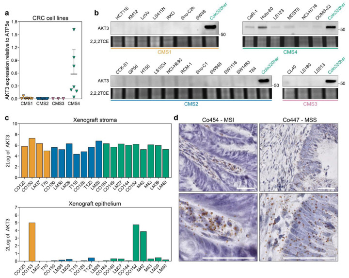
Figure 2.
AKT3 is more highly expressed in CMS4 human CRC cell lines and tumours. (a) Quantitative real time PCR analysis on 29 established CRC cancer cell lines for AKT3 expression. Expression level was normalised to ATP5E expression, average normalised expression of N = 3, each performed with technical triplicates, is plotted per cell line. Normalisation to GUSB yielded similar results; (b) Western blot analysis for AKT3 expression in the same cell line panel as in a. The CMS4 cell line Colo320-HSR was included on every blot as a positive control. Numbers on right hand side of blots indicate position of specified protein molecular weight marker. 2,2,2-Trichloroethanol (2,2,2TCE) signal (excerpt taken around 60 kDa region) indicates amount of protein loaded per cell line; (c) 2Log expression of AKT3 in the murine stroma and human cancer cells in PDX models as determined by RNAseq; (d) Representative images of RNAscope staining for AKT3 (brown dots) on human CRC tissue slides. Sections were counterstained with haematoxylin. AKT3 expression was only detected in the stroma of Co454, whereas the epithelium of Co447 also stained positive. MSI, microsatellite instable; MSS, microsatellite stable. Scale bars 50 µm.

