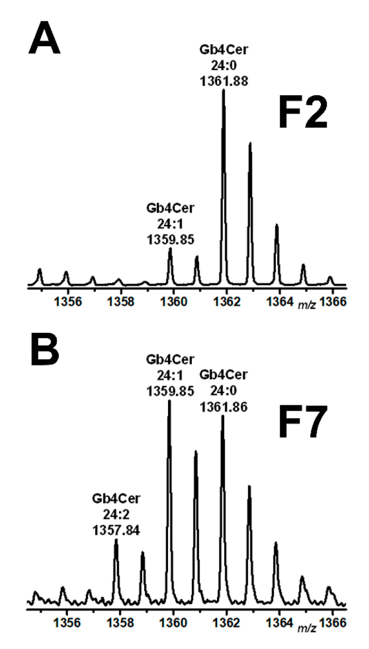
Figure 4.
Partial MS1-spectra of Gb4Cer detected in DRM fraction F2 (A) and non-DRM fraction F7 (B) obtained from a sucrose gradient of pHRCEpiCs. Gradient fractions were prepared from replicate 1 of pHRCEpiCs (see Figure 3). The spectra were recorded in the positive ion mode and span the m/z range between 1356 and 1366 showing Gb4Cer lipoforms as singly charged monosodiated [M+Na]+ species with ceramide moieties built up from a constant sphingosine (d18:1) residue and varying C24 fatty acids as indicated.

