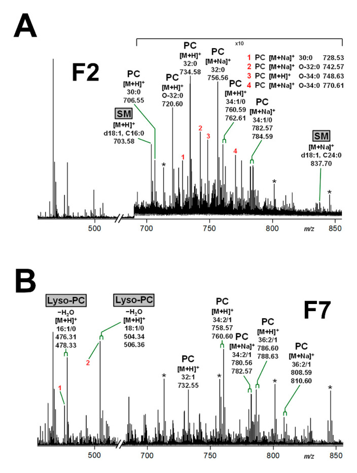
Figure 5.
MS1-spectra of phospholipids detectable in DRM fraction F2 (A) and non-DRM fraction F7 (B) obtained from a sucrose gradient of pHRCEpiCs. Gradient fractions were prepared from replicate 1 of pHRCEpiCs (see Figure 3). The mass spectra were recorded in the positive ion mode, and phospholipids were detected as singly charged protonated [M+H]+ or monosodiated [M+Na]+ species as indicated in the spectra. The exclusive presence of SM and lyso-PC species, both highlighted as grayed boxes, in the DRM fraction F2 and the non-DRM fraction F7, respectively, indicates their specific distribution to the liquid-ordered and liquid-disordered membrane phase, respectively. Possible alternative structures for [M+Na]+ species at m/z 476.31 (B, 1) and 504.34 (B, 2) are lyso-PC (O-14:0) and lyso-PC (O-16:0), respectively. The asterisks indicate polyethylene glycols (PEGs).

