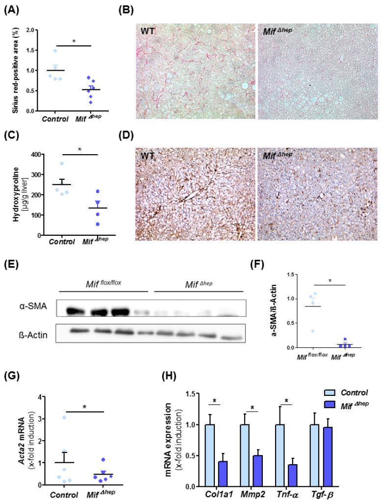
Figure 4.
Decreased liver fibrosis progression in MifΔhep mice after experimental NASH model. (A) Quantification and (B) representative Sirius red stainings of MifΔhep and control littermates mice after 8 weeks of MCD feeding. Sirius-red stainings were quantitated using ImageJ software. (C) Biochemical measurement of amino acid hydroxyproline from snap-frozen liver tissue confirmed the decreased fibrosis level within the liver. (D) Representative immunohistochemical α-SMA stainings of MifΔhep mice and control littermates after 8 weeks of MCD feeding and (E,F) Western blot analysis on whole liver tissue using antibodies against α-SMA, ß-Actin served as loading control. (F) Relative α-SMA protein level. (G) Hepatic stellate cells (HSC) activation is determined by mRNA expression level via qRT-PCR (n = 6 per group). (H) Expression patterns of fibrosis-related genes, e.g., Col1a1, Mmp2, Tnf-α, and Tgf-β were measured by qRT-PCR between MifΔhep mice and control littermates (n = 6 per group). Asterisks indicate statistical significance: * p < 0.05.

