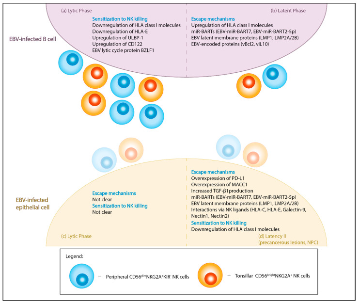
Figure 1.
Graphical summary of the interactions between NK cells and EBV-infected cells. EBV infects both B cells (a,b) and epithelial cells (c,d) in either lytic or latent forms. Peripheral CD56dimNKG2A+KIR- NK cells (blue) and tonsillar CD56brightNKG2A+ NK cells (orange) have been identified to be the main NK subset in restricting EBV-infection in B lymphocytes. The reactivity of these subsets against EBV-infected epithelial cells remains unclear. (a) During the lytic phase, EBV-infected B lymphocytes are more sensitized to NK killing due to changes in the expression of NK receptor ligands (HLA class I molecules, HLA-E, ULBP-1, CD122) and induction of lytic cycle protein BZLF1. (b) During the latent phase, there is greater NK evasion in infected B cells due to the expression of miR-BARTs, latent membrane antigens (LMP1, LMP2A/B) and other virus-encoded proteins (vBcl-2, vIL10). The miR-BARTs and LMP2A downregulate NKG2D ligands and diminish NK activation potential. The anti-apoptotic protein, vBcl2, provides protection against NK-mediated apoptosis, while pre-latent phase protein vIL-10 prevents lytic reactivation in EBV-infected tonsillar B cells. Additionally, the upregulation of HLA class I molecules can dampen NK activation through the engagement of inhibitory receptors. (c) EBV lytic infection in normal epithelial cells leads to cytopathy, while lytic reactivation in NPC results in genomic instability. However, how this affects evasion or sensitization to NK cells remains unclear. (d) In type II latency, EBV-infected epithelial cells can escape NK attack through the overexpression of PDL-1, which promote NK exhaustion and/or the overexpression of MACC1 which leads to increased TGF-β1 production. Additionally, expression of miR-BARTs, latent membrane proteins and NK inhibitory ligands (HLA-C, HLA-E), and ligation of co-inhibitory receptors on NK (Galectin-9-TIM3, Nectin1-CD96 and Nectin2-TIGIT) may also desensitize antitumor NK responses. In contrast, the downregulation of HLA class I molecules, common in NPC, may sensitize tumor cells to NK cells due to lack of NK inhibitory ligands (“missing-self”). HLA, human leukocyte antigen; ULBP-1, UL16 binding protein 1; BZLF1, BamHI Z fragment leftward open reading frame 1; BART, BamHI-A rightward transcripts; LMP, latent membrane protein; MACC1, metastasis-associated colon cancer-1; vBcl2, viral Bcl-2 homolog; vIL10, viral interleukin-10; PDL-1, programmed cell death protein ligand 1, TGF-β1, transforming growth factor-β 1; NKG2, natural killer group 2; TIM3, T-cell immunoglobulin and mucin domain 3; TIGIT, T cell immunoreceptor with immunoglobulin and ITIM domains.

