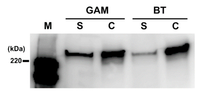
Figure 6.
Localization of Toxin A in CD culture. After CD was cultured in the conditioned medium containing PF (2.0 mg/mL) derived from GAM broth or BT, the culture was separated into the supernatant and cell pellet by centrifugation. Toxin A levels in the supernatant (S) and cell pellet (P) were evaluated by Western blotting with anti-Toxin A antibody. M, HRP-labeled protein size marker.

