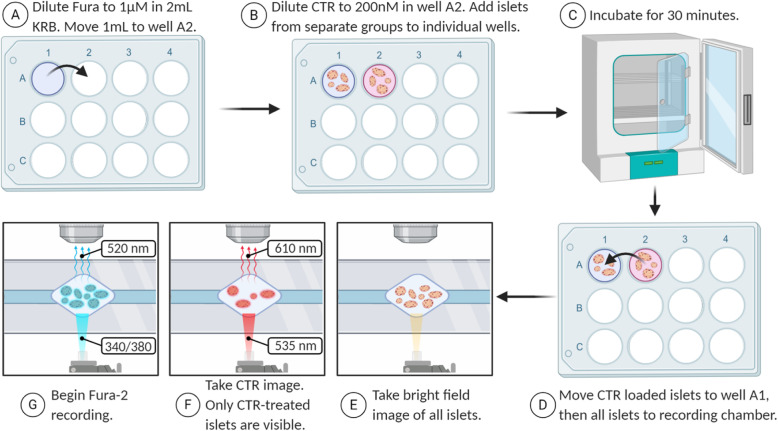
Fig. 9.
Fluorescence imaging with Fura-2 and CTR. Fura-2 is diluted in one well (a), then a 1 ml aliquot is moved to an adjacent well and CTR is added (b). Islets are added to each well and incubated for 30 min (c) before being combined in the Fura-2 only well and transferred to the recording chamber (d). Islets are imaged using three different wavelengths: bright field (e), CTR only (f), and Fura-2 (g). Once the calcium imaging experiment is complete, the islets from the second well can be identified as the red islets in the CTR image

