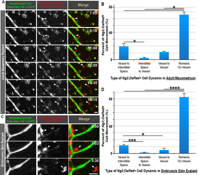
Figure 1.

Pericyte shedding during angiogenic sprouting of adult and embryonic microvessels. (A): Time-lapse images of Flk-1:eGFP+ endothelial cells (i-vi, left-most column, and green in right-most column) and Ng2:DsRed + vascular pericytes (i-vi, middle column, and red in right-most column) during pericyte detachment (arrowhead) in an adult mesentery explant model. Scale bar, 20 μm. Time (hh:mm), upper right. ‘Full movie from which nonconsecutive stills were selected, Supplemental Movie 1.’ (B): Percent of Ng2:DsRed + cell movement for each type of dynamic in an adult mesometrium explant model. Data were quantified from four biological replicates of distinct movies. *P < 0.05 by ANOVA followed by Tukey’s multiple comparisons test. Error bars, SEM. (C): Time-lapse images of Flk-1:eGFP+ endothelial cells (i-iii, left-most column, and green in right-most column) and Ng2:DsRed + vascular pericytes (i-iii, middle column, and red in right-most column) during pericyte detachment (arrowhead) in an embryonic skin explant model. Scale bar, 20 μm. Time (hh:mm), upper right. ‘Full movie from which nonconsecutive stills were selected, Supp. Movie 2.’ (D): Percent of Ng2:DsRed + cell movement for each type of dynamic in an embryonic dorsal skin explant model. Data were quantified from four biological replicates of distinct movies. *P < 0.05, ***P < 0.005, ****P < 0.001 by ANOVA followed by Tukey’s multiple comparisons test. Error bars, SEM.
