Figure 5.

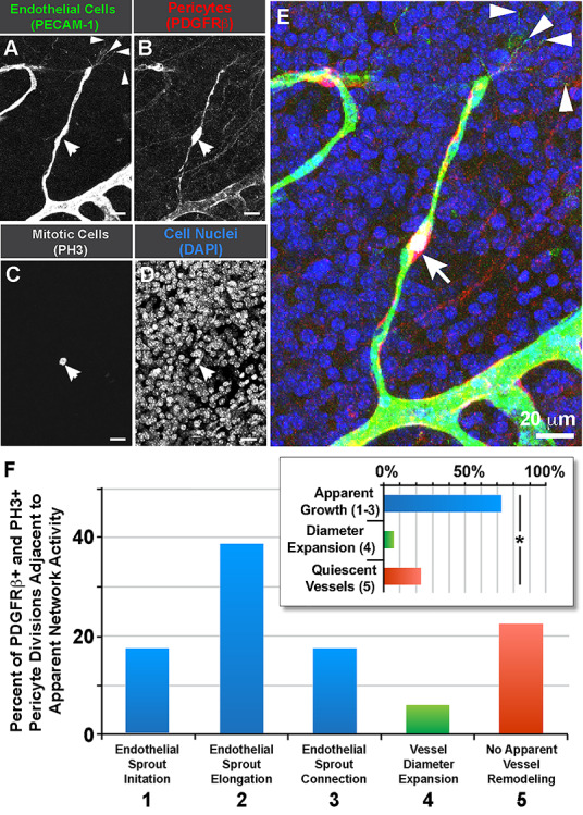
Pericyte divisions occur along endothelial sprouts in postnatal brain capillaries within the caudothalamic groove. (A): Endothelial cells labeled for PECAM-1 (green in E). (B): Vascular pericytes labeled for PDGFRβ (red in E). (C): Mitotic cell labeled for phospho-histone H3 (white in E, arrow). (D): Cell nuclei labeled by DAPI (blue in E). (E): Merged image of all signals. Endothelial ‘tip’ cell filopodia denoted with arrowheads. Scale bar, 20 μm. (F): Percent of PDGFRβ+ pericyte divisions (PH3+) located adjacent to the apparent network activity (i.e. remodeling behavior). Blue bars represent percentages of all pericyte divisions observed in regions exhibiting vessel growth behaviors (i.e. sprout initiation, elongation and connection), green bars represent percentages of pericyte divisions occurring in vessels undergoing diameter expansion, and red bars represent percentages of pericyte divisions within seemingly quiescent vessels. Inset graph: Percent of PDGFRβ+ pericytes located adjacent to the specific network activity i.e. 1–3 (blue bar) vs. 4 (green bar) vs. 5 (red bar). Data were quantified from seven distinct brain slices from two WT brains (pooled), with each pericyte as single technical event (n = 18). *P < 0.05 by chi-square analysis.
