FIGURE 2.

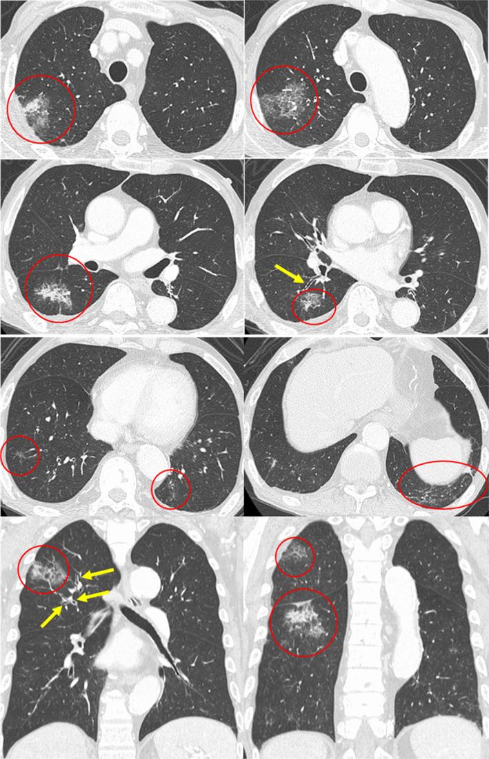
Axial and coronal chest high‐resolution computed tomography shows consolidation with peripheral ground‐glass attenuation (red circles) and thickening of bronchovascular bundles (yellow arrows)

Axial and coronal chest high‐resolution computed tomography shows consolidation with peripheral ground‐glass attenuation (red circles) and thickening of bronchovascular bundles (yellow arrows)