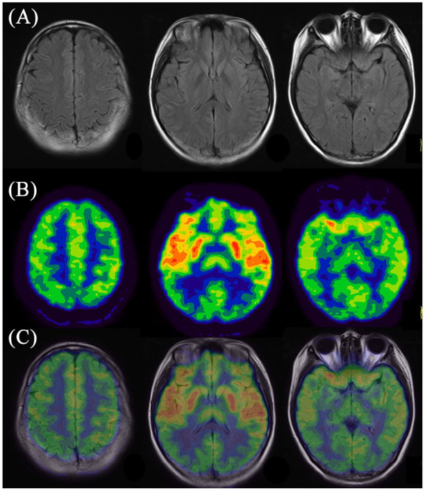
Figure 2.

Representative images of abnormal cerebral glucose metabolism in patient with paraneoplastic anti-NMDAR encephalitis detected by 18F-FDG PET imaging and corresponding MRI imaging. (A) MRI imaging; (B) 18F-FDG PET imaging; (C) PET/MRI fusion imaging.
18F-FDG PET, 18F-fluorodeoxyglucose positron emission tomography; anti-NMDAR, anti-N-methyl-D-aspartate receptor; MRI, magnetic resonance imaging.
