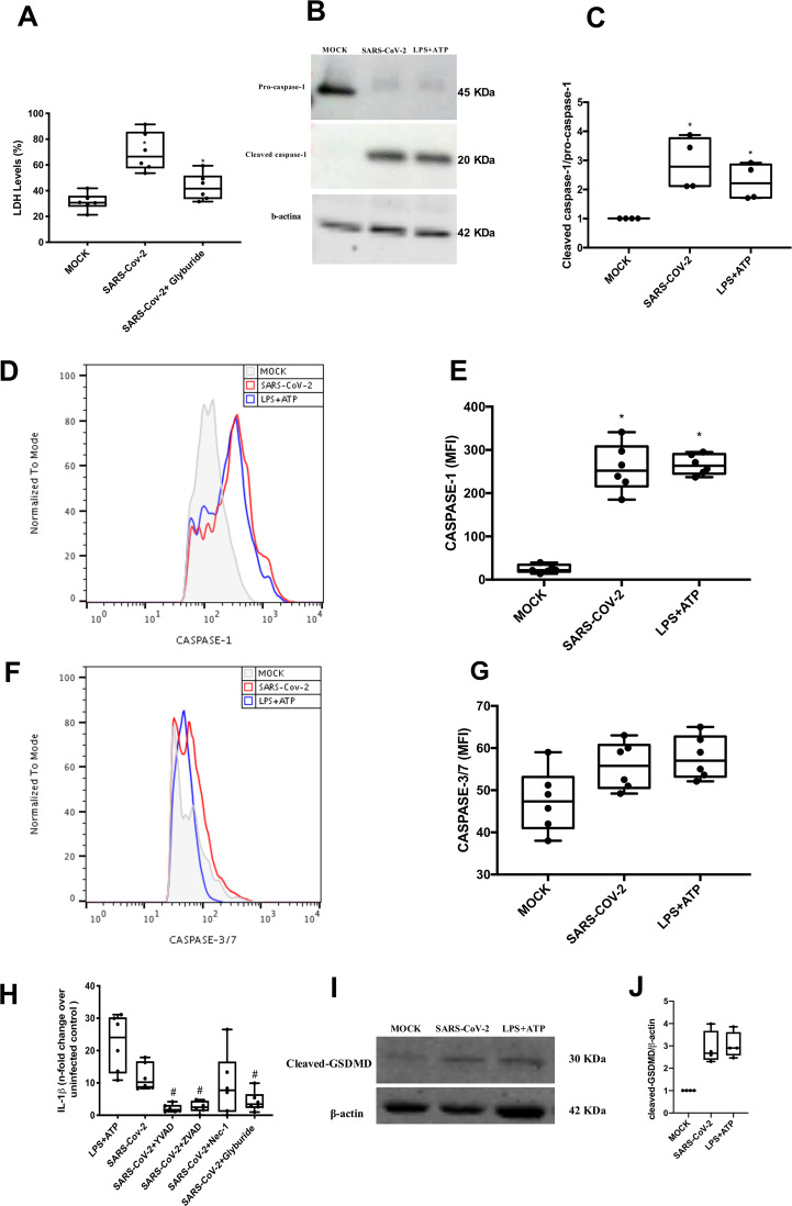
Fig. 2. Lytic cell death in SARS-CoV-2-infected monocytes associates with inflammasome engagement and pyroptosis.
Human monocytes were treated with pharmacological inhibitors to impair the function of the following proteins: NLPR3 (Gliburyde; 100 µM), caspase-1 (AC-YVAD-CMK; 1 µM), or pan-caspase (Z-VAD-FMK; 10 µM) or RIPK (Nec-1; 25 µM). Monocytes were treated since 1 h prior to infection with SARS-CoV-2 (MOI 0.1), for 24 h. As a control, monocytes were stimulated with LPS (500 ng/mL) for 23 h and, after this time, stimulated with ATP (2 mM) for 1 h. A Cell viability was assessed through the measurement of LDH levels in the supernatant of monocytes. B, C Monocytes were lysed and used for determination of pro-caspase-1 and cleaved caspase-1 levels by western blotting. D, E Monocytes were stained with FAM-YVAD-FLICA to determine the caspase-1 activity by flow cytometry. F, G Monocytes were stained with FAM-FLICA to determine caspase-3/7 activity by flow cytometry. H Cell culture supernatants were collected for the measurement of the levels of IL-1β. I, J Monocytes were lysed and cleaved GSDMD levels were determined western blotting. Western blotting images, histogram and graph data are representative of six independent experiments. Data are presented as the mean ± SEM #P < 0.05 versus infected and untreated group; *P < 0.05 versus control group (MOCK-infected).

