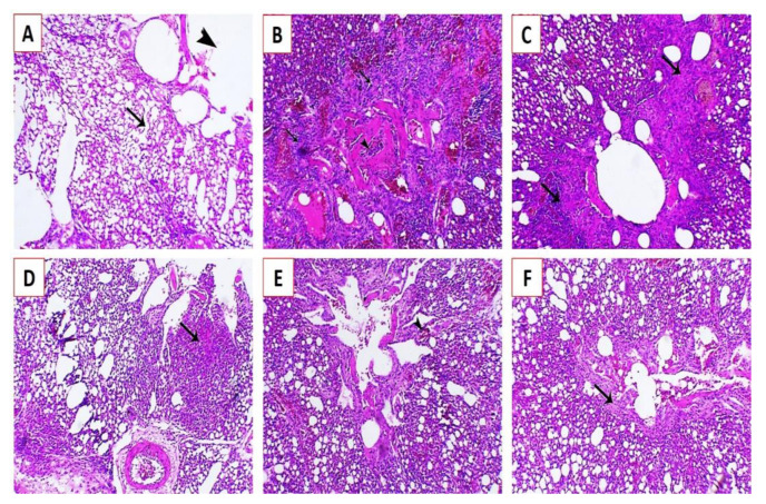
Figure 7.
Histopathological lesions in the lungs of experimentally infected Cobb chickens 5 days post-infection (dpi): (A) lung of the negative control bird (G4) showing normal parabronchi (arrowhead) and normal air capillaries (arrow); (B) lung of an experimentally infected bird in G1 showing congestion of blood capillaries and bronchial obstruction (arrowhead) attributed to peribronchial inflammatory cells infiltration (arrows); (C) lung of an experimentally infected bird in G1 showing marked endodermal hyperplasia around the parabronchi (arrows), associated with obvious inflammatory cells infiltration; (D) lung of an experimentally infected bird in G2 showing focal pneumonia associated with inflammatory cell infiltration (arrow); (E) lung of an experimentally infected bird in G2 showing mild congestion (arrowhead) and mostly patent bronchi and air capillaries; and (F) lung of an experimentally infected bird in G3 showing mild endodermal hyperplasia (arrows) and increase in the functional respiratory spaces stained by Hematoxylin and eosin (H&E X200).

