Figure 9.
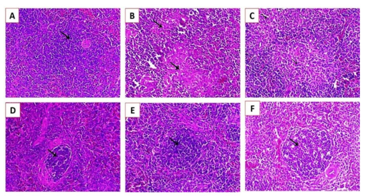
Histopathological lesions in the splenic tissues of experimentally infected Cobb chickens 5 dpi: (A) Scheme 4 showing a normal lymphoid follicle with normal lymphocytes around the central arteriole (arrow); (B) spleen of an experimentally infected bird in G1 showing marked lymphoid depletion associated with marked histocytic cell proliferation (arrows); (C) spleen of an experimentally infected bird in G2 showing marked histocytic cell proliferation (arrows); (D) spleen of an experimentally infected bird in G2 showing a normal lymphoid nodule (arrow); (E) spleen of an experimentally infected bird in G3 showing an increase in lymphoid cell proliferation within the white pulp (arrow); and (F) spleen of an experimentally infected bird in G3 showing increased lymphoid cell proliferation (arrow) (H&E, X200).

